What Is an Advance Radiology ?
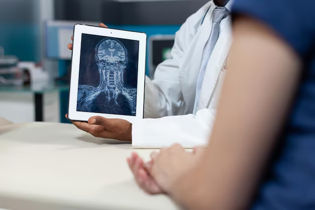
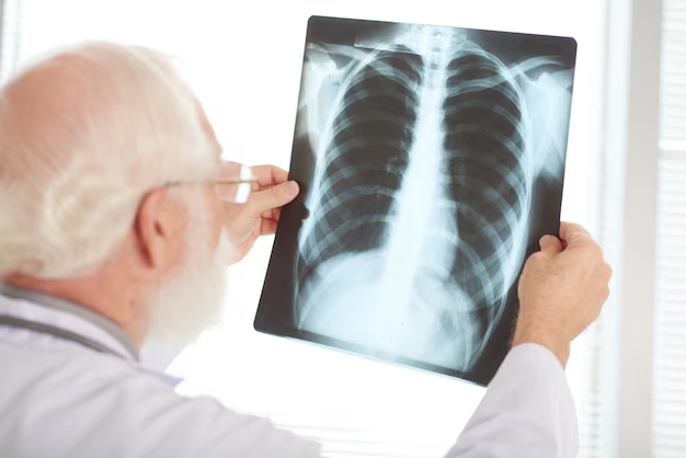
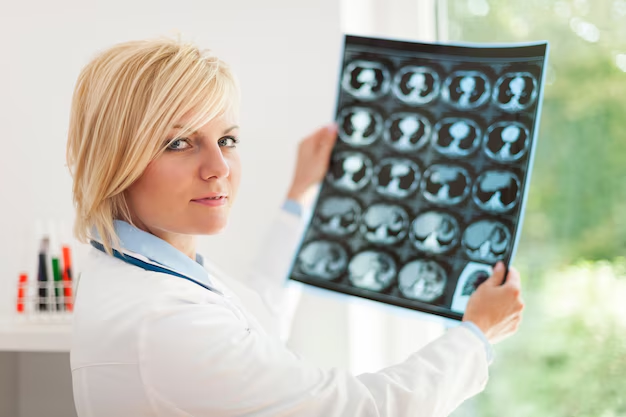
Medical imaging and radiology is the branch of healthcare which utilises advanced technologies such as CT scanners and MRI machines to provide detailed diagnosis and advanced treatment of medical conditions.Radiology may be divided into two different areas, diagnostic radiology and interventional radiology.
Radiology Services
At our Hospitals our highly experienced radiology team includes radiologists, medical physicists, radiographers and many more expertly trained healthcare professionals. We are committed to investing in the most cutting-edge technologies such as the TruBeam Radiotherapy System and 3 Tesla MRI scanners to ensure our patients receive state-of-the-art diagnostics, and as such Turkey are rightly recognised as offering some of the most innovative medical imaging and radiology services in the region.
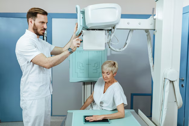
DIAGNOSTIC RADIOLOGY
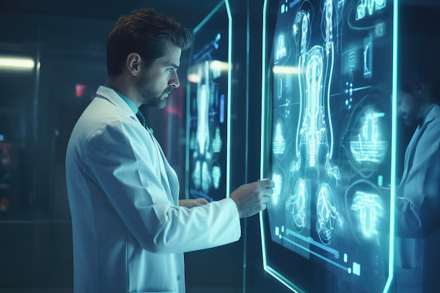
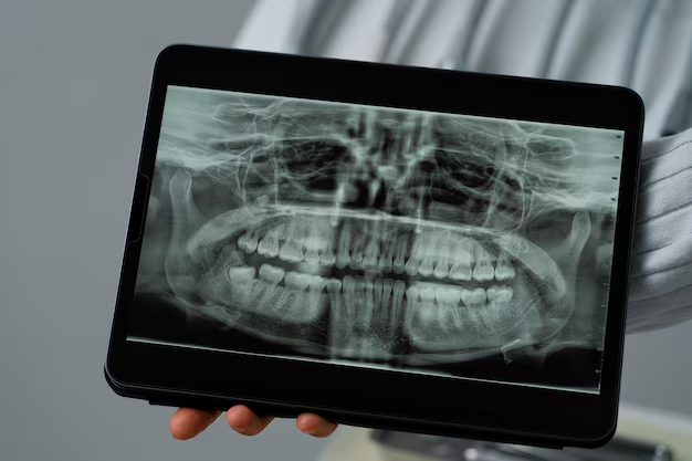
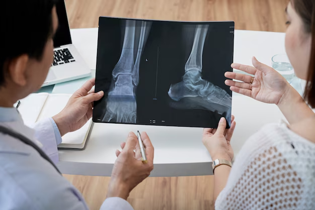
Diagnostic radiology helps health care providers see structures inside your body. Doctors that specialize in the interpretation of these images are called diagnostic radiologists. Using the diagnostic images, the radiologist or other providers can often:
- Diagnose the cause of your symptoms
- Monitor how well your body is responding to a treatment you are receiving for your disease or condition
- Screen for different illnesses, such as breast cancer, colon cancer, or heart disease
The most common types of diagnostic radiology exams include:
- Computed tomography (CT), also known as a computerized axial tomography (CAT) scan, including CT angiography
- Fluoroscopy, including upper GI and barium enema
- Magnetic resonance imaging (MRI) and magnetic resonance angiography (MRA)
- Mammography
- Nuclear medicine, which includes such tests as a bone scan, thyroid scan, and thallium cardiac stress test
- Plain x-rays, which includes chest x-ray
- Positron emission tomography, also called PET imaging, PET scan, or PET-CT when it is combined with CT
- Ultrasound
INTERVENTIONAL RADIOLOGY
Interventional radiologists are doctors that use imaging such as CT, ultrasound, MRI, and fluoroscopy to help guide procedures. The imaging is helpful to the doctor when inserting catheters, wires, and other small instruments and tools into your body. This typically allows for smaller incisions (cuts).
Providers can use this technology to detect or treat conditions in almost any part of the body instead of using larger incisions and directly looking inside of your body through a scope (camera) or with open surgery.
Interventional radiologists often are involved in treating cancers or tumors, blockages in the arteries and veins, fibroids in the uterus, back pain, liver problems, and kidney problems.
The interventional radiologist will make no incision or only a very small one. You rarely need to stay in the hospital after the procedure. Most people need only moderate sedation (medicines to help you relax).
Beard And Mustache Transplantation Stages
The stages of beard and mustache transplantation are very similar to the steps of hair transplantation on the scalp. It is as follows:
- Clinical examination: At this stage, the density of the donor area and the bald areas on the chin are defined. The number of grafts and hair follicles to be harvested is determined.
- Medical tests: Carrying out all the necessary tests, including blood and skin tests, to ensure the patient is suitable for the operation.
- Preparation for the harvesting: The donor area is shaved to harvest the hair follicles then the patient is given local anesthesia.
- Harvesting the hair follicles: The healthy hair follicles are being harvested from the donor area via a Micro Motor to be placed in the saline solution, which can preserve them until they are transplanted to the recipient area.
- Implantation stage: The follicles are placed in Choi Pens, and with only one click, the channels are opened, then the follicles are implanted simultaneously.
Examples of interventional radiology procedures include:
- Angiography or angioplasty and stent placement
- Embolization to control bleeding
- Cancer treatments including tumor embolization using chemoembolization or Y-90 radioembolization
- Tumor ablation with radiofrequency ablation, cryoablation, or microwave ablation
- Vertebroplasty and kyphoplasty
- Needle biopsies of different organs, such as the lungs and thyroid gland
- Breast biopsy, guided either by stereotactic or ultrasound techniques
- Uterine artery embolization
- Feeding tube placement
- Venous access catheter placement, such as ports and PICCs
