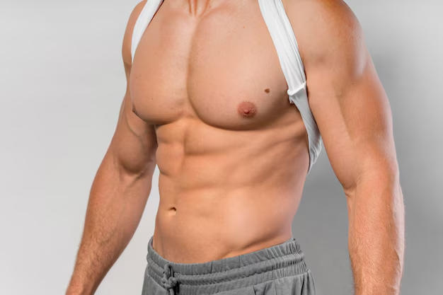
What Is Pectus Excavatum ?
Pectus excavatum (PE) also called as pigeon chest and pectus carinatum (PC) are very common chest wall deformities. Pectus carinatum is less common than pectus excavatum. Pectus excavatum is a condition in which breastbone (sternum) is sunken into chest. Pectus carinatum is a deformity in which the breastbone is pushed forward.
Pectus excavatum is often visible short after the birth. The patients may not have any symptoms during childhood. The symptoms usually become apperent during early adolescence and increase with age due to growth and decrease of the flexibility of bones. Severe cases can cause extra pressure on the heart and lungs which lead to shortness of breath, frequent respiratory tract infections, mild chest painand limited physical activities.
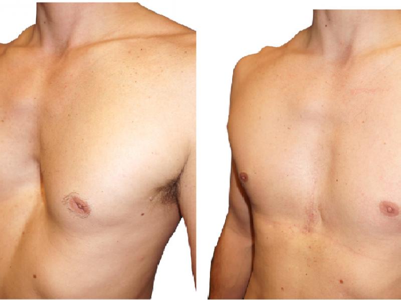
Patients who have a severe deformity and suffering from the above mentioned symptoms are good candidates for the surgery. CT scan, echocardiogram, pulmonary function tests are done before the surgery to determine the severity deformity to allow the surgeon plan the procedure. During the initial consultation to determine the most appropriate surgical options for the patients individual situation, our surgeon will provide the detailed information about the planned operation and post-operative care and the recovery process.
Video assisted minimal invasive (VATS) Nuss Procedure
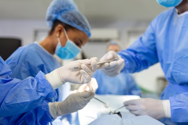
The nuss procedure is a minimally invasive surgery which involves to insert a convex bar under the ribs and sternum in order to correct the chest wall. The aim of the surgery is to correct a pectus excavatum deformity, improve the breathing and cardiac functions, and also provide a normal appearance to the chest.
The curved metal bar also called as the lorenz bar which is shaped to fit the patient is inserted through a small incision to push the sternum out to correct the deformity. When the bar is placed and the chest wall became to correct shape, a stabilizer bar is attached to the bar and fixed to the ribs to prevent the slipping. In oreder to reshape the chest wall the bar have to be remain minimum 3 years after the surgery.
NUSS PROCEDURE PECTUS EXCAVATUM
Nuss Procedure was invented by Donald Nuss in 1987. It is a minimally invasive procedure. The patient heals faster and the long term outcome of the operation is favourable. It is a widespread method in our present day.
Before the Surgery
- First, physical examination of the patient is conducted.
- The patient and patient’s relative are informed thoroughly and their questions are answered, in order for them to be able to make an informed decision about the pectus excavatum treatment.
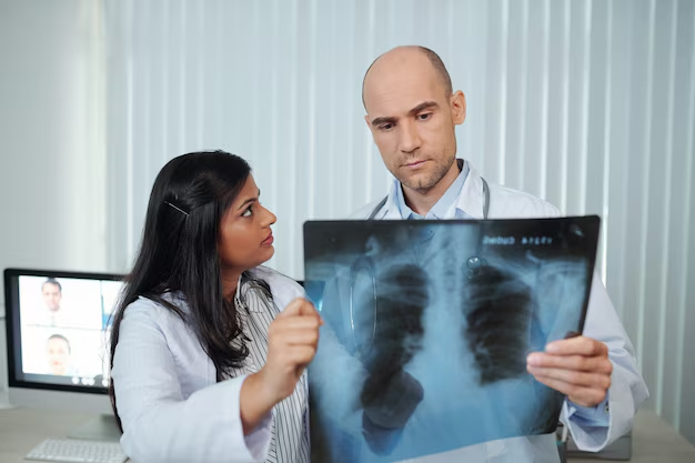
- To determine the level of chest deformation a two-sided chest graphy (front and side-back) and tomography of the chest is taken. According to Haller index, if the proportion of horizontal length of the chest to its vertical length is greater than 2.5 the case is taken into serious consideration. If the proportion is greater than 3.5 the patient may consider surgical treatment.
- Blood tests and respiratory function tests are conducted to determine whether the patient is fit for general anesthesia.
- Cardiology consultation is necessary to determine whether the patient has a heart condition. If necessary EKG and EKO tests are conducted to find a possible heart condition, if the patient indeed has a heart condition, it should be treated first in order to proceed with pectus excavatum treatment.
- For those patients with internal diseases, specific consultations are necessary.
Preparing for the Surgery
Quitting smoking at least 2 months prior to the surgery minimizes all possible problems that may arise after the surgery.
The Surgery
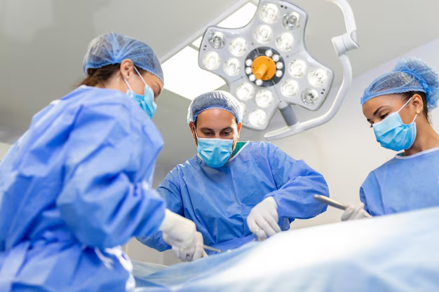
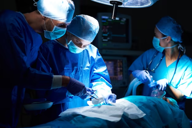
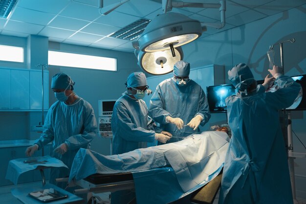
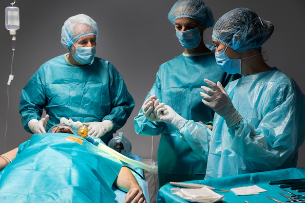
- The surgery is conducted under general anesthesia and it takes 45 – 60 minutes.
- A steel bar is implanted into the chest to pull up the sunken area.
Pectus bar is implanted into the chest through the incisions made in the thoracic wall and it is secured with muscle tissue. A metal stabilizer is also used to stabilize the bar.
Following the Surgery
The patient experiences pain in the first 3 days following the surgery. Pain is managed by epidural administration.
Generally after 3 days pain is managed by painkillers taken through the mouth or vein with injections.
Hospital stay is generally 3 – 5 days. Dressing the wound in the first 3 days is enough. The incisions may be left undressed afterwards. Rarely the patient may feel the both sides of the bar under the skin. Massaging the skin lightly prevents the bar to stick the skin. In most cases pectus bar fixes the problem without complications.
The patient can do safe exercises 3 months after the surgery. After 6 months the patient can do regular exercises such as swimming, tennis, jogging etc. Although contact sports such as wrestling, karate, bow are strictly forbidden. The patient should stay away from contact sports for the rest of his life.
What to do, what not to do after the surgery
In the first month following the surgery the patient;
- Should not bend her back (She should bend her hips).
- Should not turn back.
- Should not roll either way.
- Should not run or engage in aerobics.
- Should not engage in physically challenging activities.
In the first 3 months after the surgery the patient:
- Should not lift anything heavy (including books and back packs)
- Should not engage in aerobics.

Recommended activities
- The patient should do posture exercises in order for the pectus bar to maintain its place.
- The patient should take daily walks in order to regain her normal strength.
- The patient should do deep breathing exercises.
- The patient can do more challenging exercises at the end of 6 weeks.
- The patient should consult her doctor before starting any routine exercises.
Diet;
- The patient can go back to their normal diet as long as they take their medication.
Removing the bars
Depending on the age and the state of the thoracic wall the pectus bar is removed with a 30 minute operation conducted under general anesthesia within 2 – 4 years.
SURGICAL TREATMENT OF PECTUS CARINATUM (PIGEON CHEST) AND ABRAMSON PROCEDURE
Argentinian thoracic surgeon Dr. Horacio Abramson was the first to adapt the Nuss technique (minimally invasive technique to treat pectus carinatum patients) to treat pectus carinatum patients and share his findings in 2005 with the medical world (1).
After a five-year follow-up, he shared results acquired from 40 patients with the medical world. After the bar was removed the procedure was highly successful for 10, successful for 4, mediocre for 4, and unsuccessful for 2 out of 20 patients in total. The success rate of Abramson procedure is quite high as seen above. After the removal of the bar, results were positive for 18 out of 20 patients (2).
Before the Surgery;
- First, physical examination of the patient is conducted.
- The patient and patient’s relative is informed thoroughly and their questions are answered, in order for them to be able to make an informed decision about the pectus carinatum treatment.
- To determine the level of chest deformation a two sided chest graphy (front and side-back) and tomography of the chest is taken. According to Haller index, if the proportion of horizontal length of the chest to its vertical length is greater than 2.5 the case is taken into serious consideration. If the proportion is greater than 3.5 the patient may consider surgical treatment.
- Blood tests and respiratory function tests are conducted to determine whether the patient is fit for general anesthesia.
- Cardiology consultation is necessary to determine whether the patient has a heart condition. If necessary EKG and EKO tests are conducted to find a possible heart condition, if the patient indeed has a heart condition, it should be treated first in order to proceed with pectus excavatum treatment.
- For those patients with internal diseases specific consultations are necessary.
- The patient and patient’s relative re informed about alternative treatment methods and success rates.
- Patient’s questions about her condition and the procedure are answered prior to te operation.
Success rates of open surgery and Abramson procedure which is an endoscopic minimally invasive procedure are similar. However, Abramson procedure have become widespread due to lower chance of complications during and after the surgery, early discharge and quicker adaptation to daily life (2 weeks).
The preferred ages for the operation is 7 – 14. Between these ages bone structure of the thoracic wall is softer making the procedure less challenging. Also following the procedure thoracic wall can grow into its natural anatomical position. These being said, success rate is similar for adults (3).
Attaching the Stabilizer to the Bones
Lorenz Bar (Pectus Bar) is chosen according to the chest size and shape of the patient and the two ends of the bar are given the necessary curve before the operation. Pectus Bar is introduced into the tunnel from one end and it is pushed through until it reaches the other end of the tunnel. Once it does, the bar has reached its final position.
When the bar reaches the other side of the thoracic wall, the PVC tube is removed. Before or after the bar is inserted into the tunnel, stabilizers are placed onto the ribs with steel stitches.
Once the sternum is in the desired position, incisions are stitched up and the procedure concluded. You can get detailed information on Abramson Procedure in medical publications (4).

Removing the Bars
Depending on the patient’s age and the state of the sternum, the bar is removed within 2 – 4 years with a 30 minute procedure under general anesthesia.
Risks of the Procedure
Generally risks of the operation is very low (5). Pneumothorax (1 out of 40 patients – air leaking into the space between the chest wall and the lung), infection of wounds (1 out of 40 patients), inflammation in the scars (6 out of 40 patients), coming off of steel stitches (3 out of 40 patients). Infecting scars occurs less than 0.7% of patients (6).
Reference
- Arch Bronconeumol. 2005;41(6):349-51 and J Pediatr Surg. 2009;44(1):118-24.
- Pediatr Surg. 2009;44(1):118–24.
- http://icvts.ctsnetjournals.org/cgi/content/full/9/suppl_1/S1.
- Arch Bronconeumol. 2005; 41:349-51 ve Journal of Pediatric Surgery, 44 (1), Jan 2009, p. 118-124.
- Pediatr Surg. 2009;44(1):118-24).
- http://icvts.ctsnetjournals.org/cgi/content/full/9/suppl_1/S1.